Understanding Prostate MRI
Magnetic resonance imaging (MRI) is an imaging study that makes detailed pictures of the inside of your body. It uses radio waves, magnets, and a computer to create images of the soft tissues in the body, like the prostate gland. It does not use any kind of radiation.
An MRI can be done and used in many ways in cancer care. It can help find possible areas of cancer to decide if a biopsy is needed. It can also help guide a biopsy needle so that the changed tissue is taken out for testing.
Prostate MRI is a special kind of MRI used to look at the prostate. It can show exactly where there's changed tissue in the prostate and how large the area is. It can also show how likely it is that these changes are cancer. The images may also show whether the cancer has spread beyond the prostate to nearby lymph nodes or other tissues.
Some of the special MRIs used for the prostate are:
-
Multiparametric MRI (mpMRI). This scan might be used if screening suggests you might have prostate cancer. It uses more than one type of MRI (such as dynamic contrast-enhanced MRI, diffusion weight imaging, or MR spectrometry) to find even small tumors throughout the prostate. If cancer is found, mpMRI can help figure out how fast it's likely going to grow and spread.
-
Fusion MRI. MRIs can be used along with ultrasound to better target changes in the prostate while doing a needle biopsy.
Sometimes a dye (contrast agent) is put into your blood through a vein in your arm as part of an MRI. The dye helps get clearer details on the images. A prostate MRI might also be done with a probe that's put in your rectum. This is called an endorectal ("inside rectum") coil. The prostate is very close to the rectum. So the coil helps get certain images and can show clear details of the inside of the prostate gland.
Prostate MRI can be used both before and after a diagnosis. It may be used to help guide treatment. Prostate MRI can help your provider get an idea of how serious a cancer may be, too.
Why prostate MRI is done
A prostate MRI can be used to:
-
Help diagnose prostate cancer
-
Decide if a biopsy is needed
-
Help find changed tissue in the prostate that may be cancer
-
Get information about how quickly the cancer may grow
-
Help do a biopsy of prostate tissue (MRI targeted biopsy)
-
Show if cancer has spread to other parts of the prostate or other areas of the body
How prostate MRI is done
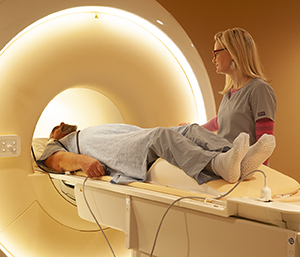
The MRI machine is a large, tube-like piece of equipment. But some newer MRIs are open on the sides. You will lie on your back on a narrow table that slides into the MRI machine. If you get contrast dye, it will be put in through an IV (intravenous) line. Then, more images will be taken. It's normal to feel a warm flush go through your body after the dye is injected.
If an endorectal coil will be used, you may be told to use an enema before the test. This helps clean out your rectum. The coil is covered and lubricated. It's then slid a short distance into your rectum. A balloon is inflated at the end of the coil inside your rectum to hold it in place during the test.
As the table moves through the machine, it takes pictures. The machine makes very loud banging and humming sounds. You'll be given headphones or earplugs to wear. The technician will be in the next room watching you through a window. You can hear and talk to the technician the whole time.
Risks of prostate MRI
There is no radiation exposure during an MRI. Still, all procedures have some risks.
A detailed safety screening will be done before your MRI. Be sure to tell your healthcare provider and the technologist if you have any implanted medical devices, metal clips or pins, or metal fragments in your body. Some devices mean you can't have an MRI because of the strong magnetic field it uses. Discuss all allergies with your healthcare team. You may not be able to have an MRI if you are allergic to contrast dye, iodine, shellfish, or certain medicines. Tell your healthcare team if you are allergic to latex, especially if your exam uses an endorectal coil. Also tell your provider if you have a serious health problem such as diabetes or kidney disease.
MRI doesn't hurt, but you will have to stay still. You may have some discomfort if an endorectal coil is used. You may feel a warming sensation in the part of your body being scanned. Let the technologist know. If dye is used, you may have an allergic reaction to it. But this is rare. Talk with your healthcare provider about any questions or concerns you have before the procedure.